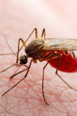
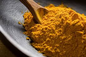
I have been doing a lot of "home remedies" right? I guess that is what fascinates me nowadays. This piece not only discusses home remedies for malaria, but also sheds insight on some other ailments, such…
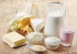
We are almost at the end of the year; unbelievable, I know. This SmartBrief is meant to give provide healthy nutrition sources to help with sustained mental and physical energy. Meals and sample menus on…
Even More Tips For Better Sex If pushups, crunches, and deadlifts aren’t your idea of a sweaty good time, you’ve still got plenty of exercise options to help keep things steamy. Pick Your Pleasure: Rather…